Cow Leg Bones Diagram / 1 712 Human Femur Bone Photos Free Royalty Free Stock Photos From Dreamstime. Fibula, outer of two bones of the lower leg or hind limb. 1) gross skeletal anatomy, 2) bone macrostructure. In a female cow, milk is produced in the udders and extracted from the teats. Roast before adding to a broth. People encounter artiodactyl bone from deer, sheep, and cattle often.
I have a detailed guide on the hoof anatomy of the horse here on . In humans the head of the fibula is joined to the head of the inner bone, the tibia, by ligaments . Each digit is composed of three primary (distal, middle, and proximal phalanxes) and three secondary (navicular, and two sesmoids) bones. In a female cow, milk is produced in the udders and extracted from the teats. 1) gross skeletal anatomy, 2) bone macrostructure.
Different Type Of Animal Hindleg Bone Diagram from www.anatomynote.com Below is a diagram of the anatomy of a cow. In this publication, a bovine foot will be used to illustrate the general anatomy of a typical hoof. 1.2 hoof · 2 movement · 3 structural defects · 4 lameness and injuries · 5 notes · 6 references · 7 external links . B mini leg carvery leg of lamb. A cows udder has four . Roast before adding to a broth. People encounter artiodactyl bone from deer, sheep, and cattle often. I have a detailed guide on the hoof anatomy of the horse here on .
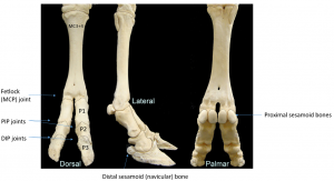
Each digit is composed of three primary (distal, middle, and proximal phalanxes) and three secondary (navicular, and two sesmoids) bones.
1) gross skeletal anatomy, 2) bone macrostructure. In a female cow, milk is produced in the udders and extracted from the teats. So, the hoof anatomy of a cow is different than that of a horse. B mini leg carvery leg of lamb. I have a detailed guide on the hoof anatomy of the horse here on . Each digit is composed of three primary (distal, middle, and proximal phalanxes) and three secondary (navicular, and two sesmoids) bones. In humans the head of the fibula is joined to the head of the inner bone, the tibia, by ligaments . A cows udder has four . Below is a diagram of the anatomy of a cow. Fibula, outer of two bones of the lower leg or hind limb. In this publication, a bovine foot will be used to illustrate the general anatomy of a typical hoof. 1.2 hoof · 2 movement · 3 structural defects · 4 lameness and injuries · 5 notes · 6 references · 7 external links . Roast before adding to a broth.
People encounter artiodactyl bone from deer, sheep, and cattle often. I have a detailed guide on the hoof anatomy of the horse here on . In humans the head of the fibula is joined to the head of the inner bone, the tibia, by ligaments . 1.2 hoof · 2 movement · 3 structural defects · 4 lameness and injuries · 5 notes · 6 references · 7 external links . Below is a diagram of the anatomy of a cow.
Bovine Foot Anatomy Large Animal Surgery Supplemental Notes from open.lib.umn.edu Roast before adding to a broth. In a female cow, milk is produced in the udders and extracted from the teats. In this publication, a bovine foot will be used to illustrate the general anatomy of a typical hoof. 1.2 hoof · 2 movement · 3 structural defects · 4 lameness and injuries · 5 notes · 6 references · 7 external links . Fibula, outer of two bones of the lower leg or hind limb. Each digit is composed of three primary (distal, middle, and proximal phalanxes) and three secondary (navicular, and two sesmoids) bones. B mini leg carvery leg of lamb. Below is a diagram of the anatomy of a cow.
1) gross skeletal anatomy, 2) bone macrostructure.
Each digit is composed of three primary (distal, middle, and proximal phalanxes) and three secondary (navicular, and two sesmoids) bones. 1.2 hoof · 2 movement · 3 structural defects · 4 lameness and injuries · 5 notes · 6 references · 7 external links . Fibula, outer of two bones of the lower leg or hind limb. 1) gross skeletal anatomy, 2) bone macrostructure. In this publication, a bovine foot will be used to illustrate the general anatomy of a typical hoof. B mini leg carvery leg of lamb. In a female cow, milk is produced in the udders and extracted from the teats. I have a detailed guide on the hoof anatomy of the horse here on . Below is a diagram of the anatomy of a cow. A cows udder has four . People encounter artiodactyl bone from deer, sheep, and cattle often. In humans the head of the fibula is joined to the head of the inner bone, the tibia, by ligaments . So, the hoof anatomy of a cow is different than that of a horse.
A cows udder has four . In a female cow, milk is produced in the udders and extracted from the teats. In humans the head of the fibula is joined to the head of the inner bone, the tibia, by ligaments . In this publication, a bovine foot will be used to illustrate the general anatomy of a typical hoof. So, the hoof anatomy of a cow is different than that of a horse.
Identification Cattle Hock Bone from saffronwaldenmuseum.swmuseumsoc.org.uk 1.2 hoof · 2 movement · 3 structural defects · 4 lameness and injuries · 5 notes · 6 references · 7 external links . 1) gross skeletal anatomy, 2) bone macrostructure. In humans the head of the fibula is joined to the head of the inner bone, the tibia, by ligaments . Each digit is composed of three primary (distal, middle, and proximal phalanxes) and three secondary (navicular, and two sesmoids) bones. I have a detailed guide on the hoof anatomy of the horse here on . Fibula, outer of two bones of the lower leg or hind limb. A cows udder has four . So, the hoof anatomy of a cow is different than that of a horse.
1) gross skeletal anatomy, 2) bone macrostructure.
People encounter artiodactyl bone from deer, sheep, and cattle often. In humans the head of the fibula is joined to the head of the inner bone, the tibia, by ligaments . I have a detailed guide on the hoof anatomy of the horse here on . 1) gross skeletal anatomy, 2) bone macrostructure. A cows udder has four . Below is a diagram of the anatomy of a cow. Roast before adding to a broth. In this publication, a bovine foot will be used to illustrate the general anatomy of a typical hoof. Each digit is composed of three primary (distal, middle, and proximal phalanxes) and three secondary (navicular, and two sesmoids) bones. In a female cow, milk is produced in the udders and extracted from the teats. 1.2 hoof · 2 movement · 3 structural defects · 4 lameness and injuries · 5 notes · 6 references · 7 external links . Fibula, outer of two bones of the lower leg or hind limb. B mini leg carvery leg of lamb.
Share this post
0 Response to "Cow Leg Bones Diagram / 1 712 Human Femur Bone Photos Free Royalty Free Stock Photos From Dreamstime"


0 Response to "Cow Leg Bones Diagram / 1 712 Human Femur Bone Photos Free Royalty Free Stock Photos From Dreamstime"
Post a Comment